
Fig. 2.
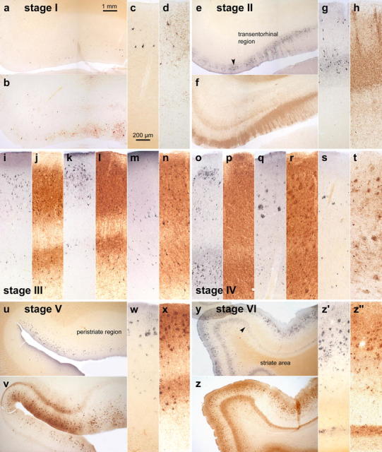
Comparison of Gallyas silver- and AT8-immunostaining of cortical neurofibrillary pathology as seen in adjacent serial 100 μm polyethylene glycol-embedded sections. The distribution pattern of the lesions throughout the various cortical fields that are necessary for staging purposes basically corresponds in both methods. It is possible with either technique to assess the progress of the neurofibrillary pathology. In the revised staging procedure, however, the greater emphasis on the presence of abnormal plexuses, which also include non-argyrophilic pretangle material in AT8-ir sections, facilitates rapid diagnostic assessment of the stages. a–d stage I: Mild involvement is confined to the transentorhinal region. Note that the plexus of AT8-ir nerve cell processes (b and d) is more conspicuous than that of argyrophilic neuropil threads (a and c). Sections originate from a non-demented 62-year-old male. e–h stage II: Lesional density increases and the pathology extends into the entorhinal region. Layer pre-α gradually sinks into a deeper position at the border between entorhinal and transentorhinal region (arrowhead). Note the greater breadth of the ir-plexus in comparison to silverstained nerve cell processes (compare f and h with e and g). Immunoreactions begin to show the deep entorhinal plexus (pri-α). The sections were obtained from a non-demented 78-year-old male. i–n stage III: The pathology in the outer and inner entorhinal (i, j) and transentorhinal (k, l) cellular layers worsens, and lesions extend into the adjoining neocortical association areas of the fusiform (occipito-temporal) gyrus (m, n). The sections originate from an 85-year-old female. o–t stage IV: The density of the lesions increases in both the entorhinal region (o, p) and fusiform gyrus (q–r) with a gradual decrease of the pallid lines (lamina dissecans in p and outer line of Baillarger in r). The neurofibrillary pathology now extends up to the medial temporal gyrus (s, t). Sections were taken from an 80-year-old female. u–x stage V: The lesions extend widely into the occipital lobe and appear in the peristriate region. Note the presence of a deep plexus in AT8-immunoreactions (v, x). Sections were obtained from a 66-year-old demented female. y-z′ stage VI: Lesions are visible even in the parastriate and striate areas of the occipital neocortex. Note the clear-cut line in layer V of the striate area (z′ and z″). The sections originate from a demented 75-year-old male. Scale bar in a applies to all overviews and that in c to all micrographs of cortical areas