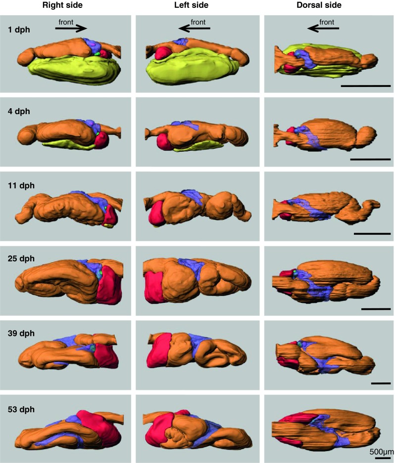
Fig. 4.

Ontogeny of the digestive organs in Atlantic cod larvae. 3D models were reconstructed from serial histological sections using Imaris software. The digestive organs are shown from three angles; right, left, and dorsal side. Arrows show the direction of the front (mouth). Orange outer layer of digestive tract, red liver, green gallbladder, purple pancreas, pink islet of Langerhans, yellow yolk-sac. Transparent colour is used for exocrine pancreas (purple) in order to show islets of Langerhans (pink) and gallbladder (green)
