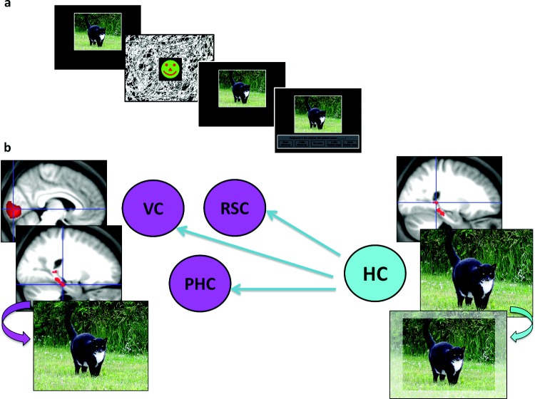
Figure 6.
An functional magnetic resonance imaging study of boundary extension (BE; Chadwick et al., in press). (a) A modified version of the task described in Figure 4 was used with healthy participants. The analysis focused exclusively on the presentation of the first scene and contrasted trials in which the BE error was later committed with those where it was not committed. (b) This revealed activity in the hippocampus (HC) and parahippocampal cortex (PHC). Connectivity analyses showed that the HC seemed to drive the BE effect, exerting top-down influence on PHC, retrosplenial cortex (RSC), and as far back down the processing stream as early visual cortex (VC).

