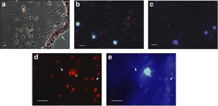
Figure 5.
Epifluorescent micrographs of CARD-FISH-labeled bacteria within the C. intestinalis tunic. (a) Gram-stained tunic and cuticle section. White arrow denotes exterior cuticle. (b) C. intestinalis individual 5 from Woods Hole July 2011 collection. EubI-III-labeled bacteria (red), Euk516-labeled tunicate cells (green) and DAPI-stained DNA (blue). (c) Same tunicate individual as in a with double Non338 (red, green) negative control showing no hybridization. DNA is stained with DAPI (blue). (d) C. intestinalis individual 2 from Woods Hole December 2011 collection. CF319a-labeled Flavobacteria-Cytophaga bacteria (red). Unlabeled bacteria are indicated with white arrowheads. White arrow shows tunicate cell. (e) Same image as C showing DAPI-stained DNA (blue). White scale bars are 10 μm.

