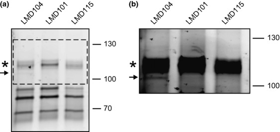
Figure 3.

Covalent binding of the fluorescent penicillin derivative Bocillin FL to PBPs present in sporulating Bacillus subtilis cells. (a) Membranes from strains LMD104 (wild-type SpoVD-mCherry; positive control), LMD101 (SpoVD deletion mutant; negative control) and LMD115 (active-site SpoVD-mCherry mutant). Cells were sporulated by resuspension at 30 °C, and membranes were isolated from cells at 5 h of sporulation (see Fig. 2). After incubation with Bocillin FL, membrane proteins were separated by 8% SDS-PAGE. The electrophoresis was run to resolve high-molecular-weight proteins. The gel was analyzed by fluorometry. (b) shows an overexposure of the area outlined by the dashed box in panel (a) in order to reveal the relatively weak fluorescence intensity of the band corresponding to SpoVD-mCherry in LMD104. The position of Bocillin FL-labeled SpoVD-mCherry is indicated by an arrow. An asterisk indicates the position of PBP1a/b identified on the basis of published data (McPherson et al., 2001). Molecular mass markers, in kDa, are indicated on the right-hand side of the panels.
