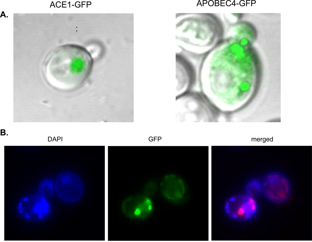
Fig. 6.
Intracellular distribution of GFP-fused deaminases in yeast. a) Confocal LSM images of cells of BY4742 ung1 producing nuclear ACE1-GFP or APOBEC4-GFP. Live cells were imaged at UNMC core Confocal microscopy facility by Zeiss LSM 410 confocal laser microscope. b) Intracellular distribution of hAIDSc-GFP in BY4742 ung1. Cells were imaged with Olympus 100 × 1.35 NA oil immersion objective on the Delta Vision microscopy system (Applied Precision, USA) consisting of an Olympus IX70 microscope (Olympus America, USA) and a CoolSnap HQ 12-bit camera (Photometrics/Roper Scientific, USA) and controlled by SoftWoRx software.

