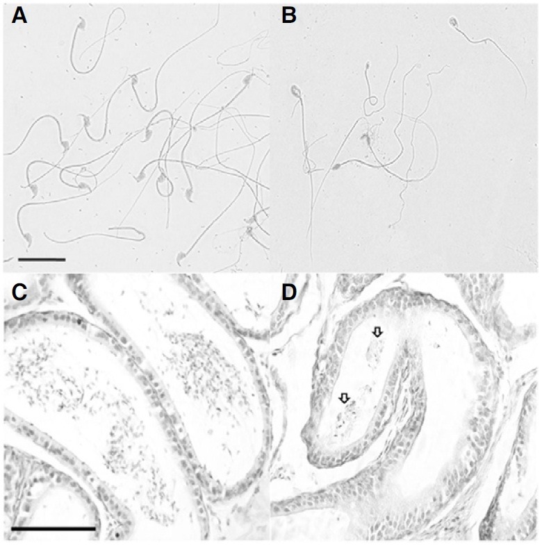
Fig. 1. Histological analysis of cauda epididymis and abnormally shaped testicular spermatozoa in 5-month-old pcd3J-/- mice. Spermatozoa from wild-type littermates (A) and from pcd3J-/- mice (B) with severely malformed heads and disorganized tails of uneven thickness. The cauda epididymis from pcd3J-/- mice (D) contained few sperm compared to the wild-type controls (C). Scale bars: 20 μm in panels A and B; 100 μm in panels C and D.

