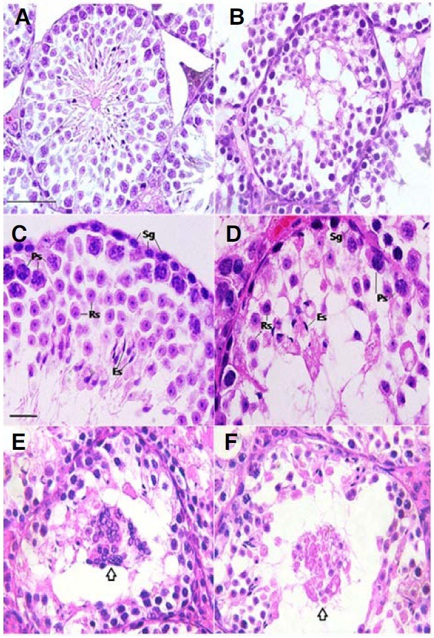
Fig. 3. Histological analysis of testes from adult pcd3J-/- mice. The testicular sections of wild-type (A, C) and pcd3J-/- (B, D, E and F) mice at postnatal day 60 were stained with hematoxylin and eosin. (A, B) there are significantly fewer differentiating male germ cells in pcd3J-/- mice compared to wild-type controls. (C, D) pachytene spermatocytes, round spermatids and elongated spermatids were present, but in significantly lower numbers in testes from pcd3J-/- mice compared to wild-type controls (Sg, spermatogonia; Ps, pachytene spermatocyte; Rs, round spermatid; Es, elongated spermatid). (E, F) Aggregated haploid cells (arrows) are present and appear to be degenerating within the lumen of the seminiferous tubules of pcd3J-/- mice. Scale bars: 100 μm in (A, B) 20 μm in (C-F).

