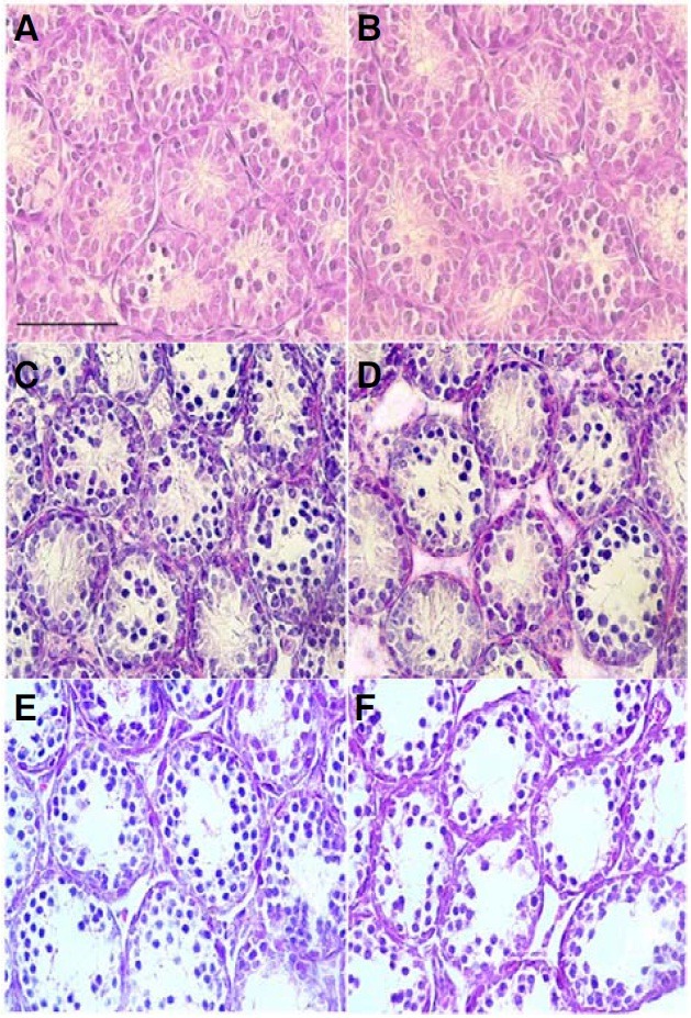
Fig. 5. Histological analysis of postnatal testicular development in pcd3J-/- mice. Representative histological sections of testes from wild-type (A, C, and E) and pcd3J-/- mice (B, D, and F) at postnatal days 12 (A, B), 15 (C, D) and 18 (E, F) stained with hematoxylin and eosin. Tissue from three animals was analyzed at each developmental stage. A decrease in the number of germ cells within the seminiferous tubules of the pcd3J-/- mice was apparent at postnatal day 18 (E, F). Scale bars: 100 μm.

