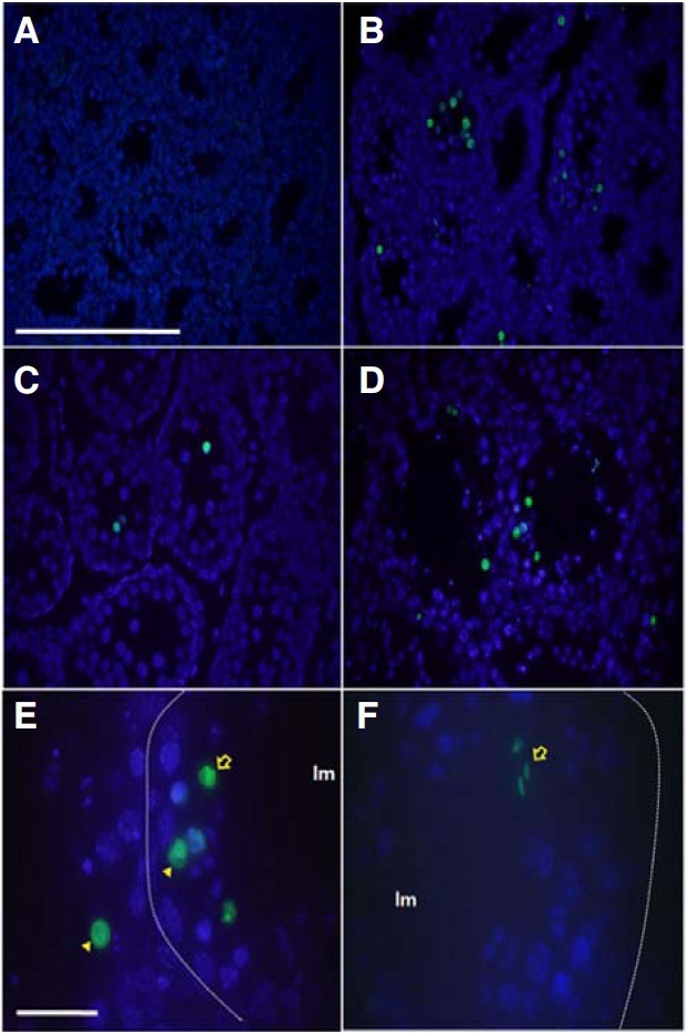
Fig. 6. Identification of apoptotic cells in the testes of pcd3J-/- mice during spermatogenesis using TUNEL staining. Representative TUNEL reactions from the testicular sections of three pcd3J-/- mice at postnatal days 12 (A), 15 (B) and 18 (C), and at 4 months of age (D-F). TUNEL-positive cells are indicated in green and counterstained with DAPI. (A) Lack of apoptotic signals at postnatal day 12. (B-D) An increase in apoptotic-positive cells from postnatal day 15 onwards. (E) A higher magnification of the seminiferous tubule reveals TUNEL-positive signals in pachytene spermatocytes (arrow-head) and round spermatids (arrow). The position of the basal lamina is indicated by the dotted lines. The lumen is indicated by lm. (F) TUNEL-positive elongated spermatids (arrow). Most of cells from the wild type TUNEL positive control experiment were TUNEL positive (data not shown). Scale bars: 100 μm in (A-D) and 20 μm in (E, F).

