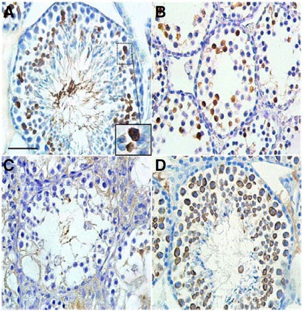
Fig. 7. Immunohistochemical localization of AGTPBP1 in mouse testes. Testes sections from wild-type (A, B) and pcd3J-/- (C) mice at 4 months of age (A) or at postnatal day 18 (B) were stained with a human anti-AGTPBP antibody. (A) Germ cells between late stage primary spermatocytes and round spermatids were positive for the antibody staining. The image within the large square is the enlarged image of the small square, indicating the staining of pachytene spermatocytes. (B) Late stage spermatocytes were positive for the antibody. (C) No clear expression of AGTPBP1 was observed. (D) In the wild type testis, all spermatogenic cells from the spermatocyte to round spermatid stages were stained by the anti-MVH antibody as a positive control for developing germ cells. Scale bars, 100 μm for (A-D).

