Figure 2.
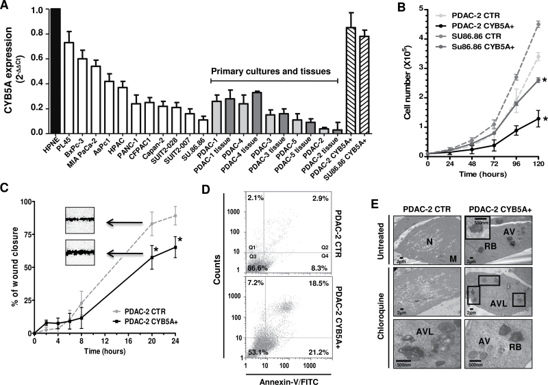
Effects of CYB5A modulation on pancreatic cells. A) CYB5A mRNA expression in pancreatic cancer cells (vs normal pancreatic ductal cells HPNE), including the corresponding tissues for five primary cell cultures (gray bars) and cells transduced with the CYB5A vector (CYB5A+). B) Growth curves of SU.86.86 and PDAC-2 cells (CTR vs CYB5A+). C) In vitro migration of CTR vs CYB5A+ cells, showing statistically significant wound healing (representative pictures in the inserts). Statistically significant differences were already detected at 8-hour time points for SU.86.86 (−30% migration vs CTR, data not shown). D) Histograms of cytofluorometric analysis of apoptosis 24 hours after starvation, as detected by Annexin-V (Q2: late apoptosis; Q4: early apoptosis) in PDAC-2 cells. E) Electron microscopy phenotypes in PDAC-2 and SU.86.86 cells, showing autophagic vacuoles (AV), residual bodies (RB), and autophagolysosomes (AVL) near the nucleus (N) and mitochondria (M). Points indicate mean values from three independent experiments. Bars indicate standard deviation. All P values were calculated with two-sided Student t test. *P < .05.

