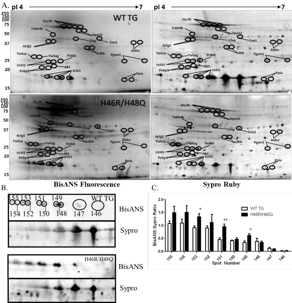
Figure 1.
Altered surface hydrophobicity of mutant SOD1 and non-SOD1 proteins in the spinal cords of symptomatic ALS mice. A) Representative 2D gels of wild type transgenic human SOD1 (WT TG, n = 8) and H46R/H48Q (n = 12) with molecular weights (left axis) and isoelectric points (pI, upper axis). Spots that significantly differed from WT TG in hydrophobic ratio are circled and annotated based on the gene names of their accession numbers identified by MALDI-TOF mass spectrometry. B) Enhanced region of 2D gels containing WT SOD1 and H46R/H48Q mutant SOD1 proteins. BisANS fluorescence and corresponding total protein stained with Sypro Ruby are shown. Quantitated SOD1 spots are shown with numbered ellipses and correspond to the quantitated hydrophobic ratio shown in C). Bars represent the mean hydrophobic ratio +/- standard deviation of 10- 8 mice per group. *p<0.05, **p<0.01 by one-way ANOVA.

