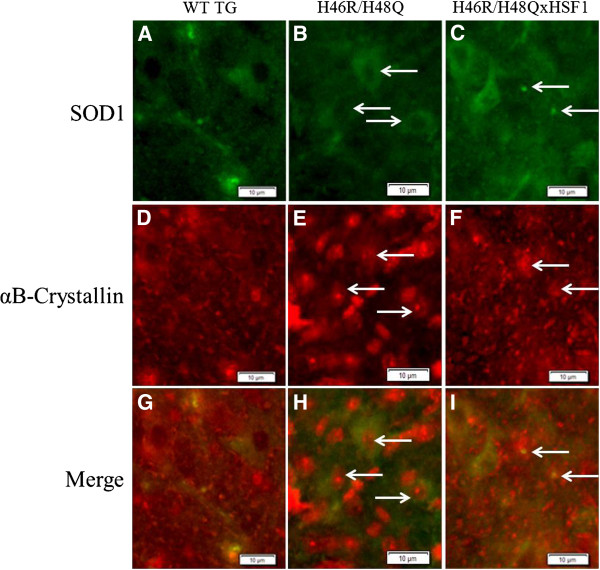
Figure 8.
Anterior horn region of lumbar spinal cord sections from mice at 220 days. SOD1 staining (A-C) and small SOD1 positive punctae in H46R/H48Q vs. H46R/H48QxHSF1 tissues (C, arrows) co-localized diffusely with αB-crystallin throughout WT TG and H46R/H48QxHSF1 tissues (D,F). Strikingly, αB-crystallin staining appeared more punctate and localized to cell nuclei of SOD1 expressing cells (E,H, arrows and G-I) in H46R/H48Q tissues. Scale bar represents 10μm.

