
Figure 4.
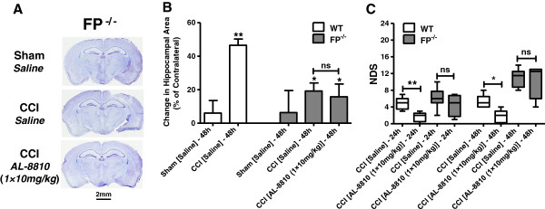
The effects of FP receptor deletion on the anatomical and behavioral outcomes after controlled cortical injury (CCI). (A) Representative photographs of cresyl violet-stained brain sections between 1 and 2 mm posterior from bregma from sham- and CCI-injured FP-/- mice 48 hours after injury. (B) Quantitative analysis of related hippocampal areas normalized to contralateral side in sham, and saline- and AL-8810- (10 mg/kg) treated mice that underwent CCI. Data are presented as mean ± SEM, **P < 0.01, versus saline-treated WT sham group, and #P < 0.05, versus saline-treated WT CCI group. ‘ns’ denotes not statistically different, one-way ANOVA followed by Tukey's multiple comparison test (n = 4 to 9). (C) Neurological deficit scores (NDS) in CCI-injured FP-/- mice in comparison with WT mice 24 and 48 hours after injury. Post-treatment with AL-8810 (10 mg/kg) had no significant effect on NDS in CCI-injured saline or AL-8810-treated FP-/- mice; there was also no significant difference between FP-/- and WT mice who underwent CCI at both time points. *P < 0.05, **P < 0.01. ‘ns’ denotes not statistically different, Kruskal-Wallis ANOVA on ranks followed by Dunn's multiple comparison test (n = 4 to 6).