
Figure 5.
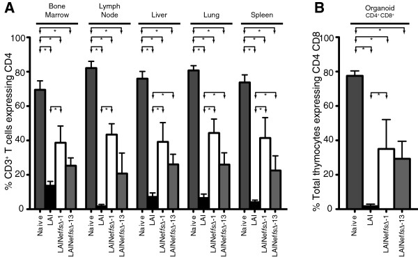
Analysis of CD4+ T cells from tissues in mice exposed to LAI, LAINeffsΔ - 1 or LAINeffsΔ - 13. (A) Percent CD4+ T cells of total T cells in five organs from un-exposed BLT mice (Naive, n = 8) were compared to groups of BLT mice exposed to one of three viruses: LAI (n = 6), LAINeffs ∆-1 (n = 4), or LAINeffs ∆-13 (n = 4). Statistical comparisons reaching significance are indicated by lines and arrows above respective bars (*, p < 0.05). (B) The same groups as in (A) were compared for CD4+CD8+ double positive thymocytes relative to total thymocytes.