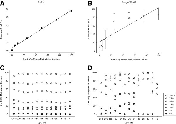
Figure 3.
Bisulfite amplicon sequencing (BSAS) and Sanger/epigenetic sequencing methylation analysis (ESME) methylation quantitation of whole genome mouse methylation controls. (A,B) Standard curves were generated by BSAS and Sanger methylation quantitation and plotted out with expected percentage of methylation versus the actual quantified methylation. Points represent the mean of the 13 CpG sites analyzed in the Rho promoter from each methylation standard (n = 3/control). Error bars = SD. (C,D) Average measured methylation for each standard plotted across each CpG site from BSAS and Sanger methylation quantitation (error bars not included for legibility).

