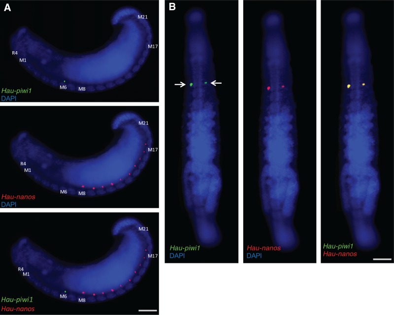
Fig. 6.
Differential expression of Hau-nanos and Hau-piwi1 in the male and female PGCs, respectively. Two color fluorescent in situs for Hau-piwi1 (green) and Hau-nanos (red). (A) Lateral views of early stage 11 embryo; Hau-piwi1 is expressed in the female PGCs of segment M6 (top), Hau-nanos is expressed in male PGCs of segments M8–M17 (center) and there is no overlapping expression (bottom). (B) Ventral views of late stage 11 embryo; by this time, Hau-piwi1 (left) and Hau-nanos (center) are coexpressed in the female PGCs (right); Hau-nanos expression is not detected in the male germline at this stage. Scale bars, 100 µm in (A); 200 µm in (B).

