Fig. 7.
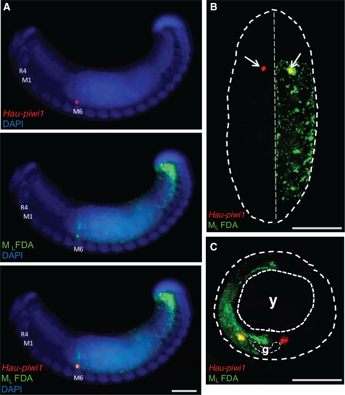
Female PGCs originate from segmental mesodermal blast cell sm10. (A) Fluorescence micrographs of embryos in which the left mesodermal teloblast (ML) was injected with FDA (green) 15 cell cycles after its birth, i.e., after the M teloblasts had each generated six presegmental blast cells (em1–em6) and nine segmental blast cells (sm1–sm9). The embryos were examined by confocal microscopy at stage 11, after processing by ISH with probes for Hau-piwi1 (red). The piwi-expressing cell(s) in segment M6 were labeled with lineage tracer on the left side. (A) Lateral view showing left side, anterior to left. (B) Ventral view, anterior up showing roughly eight segments. Dotted contour outlines the visible portion of the embryo; on the left (apparent right) side, the piwi-positive cluster is in the first labeled segment, indicating that the female PGCs originate from blast cell sm10 in the M lineage. (C) Transverse section of a similar embryo. Coarse dotted contour outlines the embryo; finer contours outline the yolk (y) and the ventral ganglion (g), Scale bar, 100 µm in (A); 50 µm in (B); 30 µm in (C).

