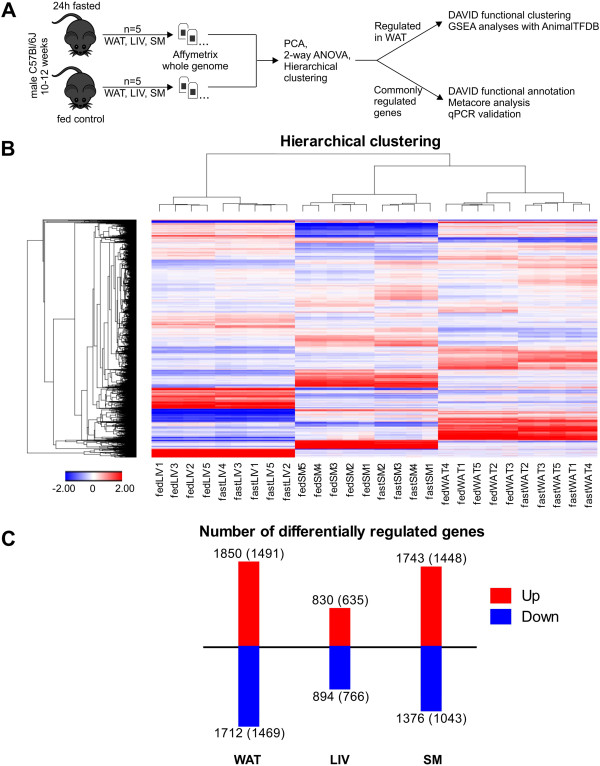
Figure 2.
Overview of microarray experiments in WAT, LIV, and SM 24 hours after onset of fasting. (A) Experimental design of the transcriptome study (WAT = white adipose tissue, LIV = liver, SM = skeletal muscle, PCA = principal component analysis). (B) The heatmap contains about 7000 probe sets differentially expressed ≥ 1.3-fold (FDR5) between fasted and fed in at least one condition. Hierarchical clustering of experiments clusters biological replicates together. Two outlier replicates were identified by principal component analysis and removed from the data set. (C) Numbers of up- and downregulated (1.3x, FDR5) microarray probe sets and RefSeq-annotated genes (in parentheses) in WAT, LIV, and SM of mice fasted for 24 hours. Additional file 1 provides detailed expression values for these genes.

