Fig. 2.
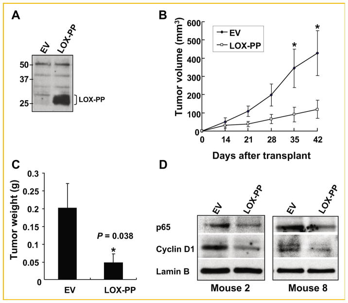
LOX-PP suppresses pancreatic tumor formation by MIA PaCa-2 cells in vivo. MIA PaCa-2 cells infected with CXbsr-EV or CXbsr-LOX-PP viruses were subcutaneously injected (4 × 10⁶ cells per injection) in both flanks (EV, left; LOX-PP, right) of NCrnu/nu nude mice (n = 8). A: Expression of LOX-PP was confirmed by immunoprecipitation and immunoblotting as described in the Materials and Methods Section before injection of cells. B: Tumor volumes were measured as described in the Materials and Methods Section and plotted as a function of days after injection (transplant). Bars represent SEM; *statistically significant differences. C: Average tumor weights were determined on day 42. Bars represent SEM (P = 0.038). D: Nuclear extracts of tumors of MIA PaCa-2-LOX-PP and MIA PaCa-2-EV xenografts from Mouse 2 and 8 were analyzed by immunoblotting for expression of the NF-κB subunit p65, cyclin D1, and Lamin B.

