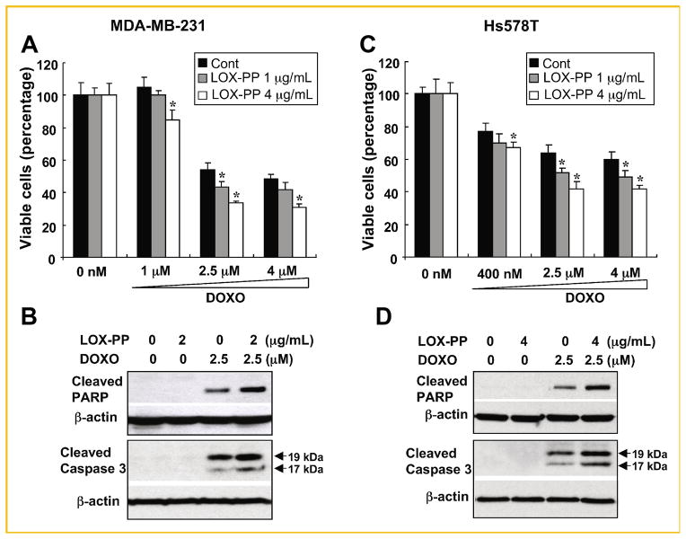
Fig. 6.
LOX-PP enhances doxorubicin-induced apoptosis in human ER-negative MDA-MB-231 and Hs578T breast cancer cells. A, C: Cells were pre-treated with 1 or 4 μg/ml LOX-PP for 24 h and then with the indicated doses of doxorubicin for an additional 24 h. Cell viability was determined by measuring ATP production. Data represent mean ± SD of quadruplicates. P values were calculated using Student’s t-test, *P <0.05. B, D: MDA-MB-231 and Hs578T breast cancer cells were treated with the indicated amounts of LOX-PP and doxorubicin (2.5 μM) individually or in combination as described in Figure 3C and WCEs analyzed by immunoblotting for cleaved PARP and caspase 3 expression, and β-actin for loading control.

