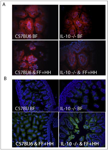
Figure 4.
Fluorescence microscopy and immunohistochemistry of intestinal samples. (A) Tight junction staining, JAM-1 localization. Top left: WT BF mouse pups. Top right: IL-10 −/− BF pups. Bottom left: WT FF + HH mouse pups. Bottom right: IL-10 −/− FF+HH mouse pups. (B) iNOS localization fluorescein isothiocyanate (FITC). Top left: WT BF mouse pups. Top right: IL-10 −/− BF pups. Bottom left: WT FF + HH mouse pups. Bottom right: IL-10 −/− FF+HH mouse pups. Also shown are DAPI-stained nuclei.

