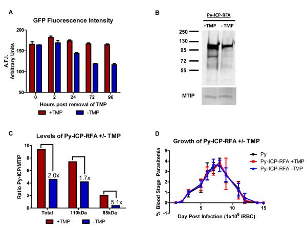
Figure 8. Destabilization reduces Py-ICP levels but does not affect blood stage growth in vivo.
(A) Monitoring de-stabilization of Py-ICP-RFA by GFP fluorescence intensity. SW mice on TMP were infected with Py-ICP-RFA parasites and parasites were allowed to propagate for 48hrs at which point TMP was removed from a subset of mice (Time 0). Blood was collected from mice +/− TMP at 0-, 2-, 24-, 72-, and 96- hrs following removal of drug and the average fluorescent intensity (A.F.I.) of GFP from 5 × 10 5 infected RBCs was determined using a fluorescence plate reader. A.F.I. is expressed as arbitrary units. (B) Representative western blot analysis of Py-ICP BS parasite lysates from mice treated with (+TMP) or without TMP (−TMP). ICP expression was detected by anti-HA antibody. The gel was also probed with a rabbit polyclonal antibody against the myosin tail interacting protein (MTIP) (Bergman et al., 2003) for loading control. (C) Bar graph depicting the densitometric ratio of ICP/MTIP from the western blot. Ratios of either total, un-processed (110kDa), or processed (85kDa) protein are depicted. Numbers indicate the fold reduction of protein following removal of TMP. These results are representative of 3 independent experiments. (D) Reduction of ICP levels does not impact blood stage replication. BALB/cJ mice were infected with Py or Py-ICP-RFA parasites in the presence or absence of TMP and blood stage parasitemia was monitored via Geimsa staining of peripheral blood smears. Py-ICP-RFA parasites had a similar growth rate and parasitemia to WT Py regardless of whether the Py-ICP-RFA was being stabilized by TMP.

