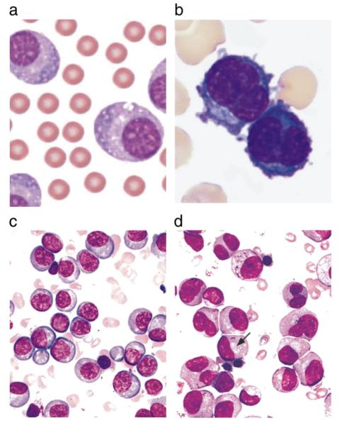
Fig. 1.
a and b. Peripheral blood smear showing the morphology of patient’s leukemic cells. Chan et al. case report.51 c and d. Bone marrow aspirate smears from two different patients with multiple myeloma, illustrating a preponderance of mostly mature-appearing plasma cells with eccentrically placed nuclei and prominent Golgi zones (arrow) (Wright Giemsa stain). From Brunning, RD, McKenna, RW. Tumors of the bone marrow. Atlas of tumor pathology (electronic fascicle), Third series, fascicle 9, 1994, Washington, DC. Armed Forces Institute of Pathology.

