
FIGURE 1:
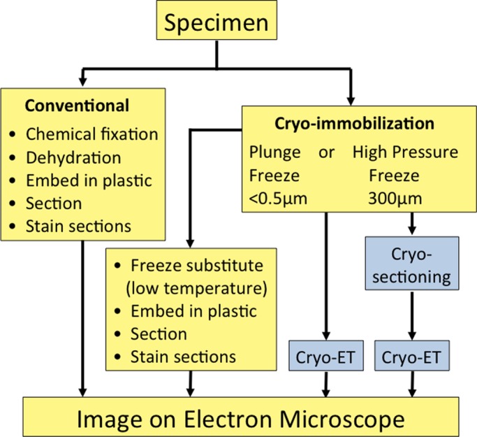
A brief flowchart showing the work to be done with different types of sample preparation for conventional electron microscopy (yellow background). The advanced cryo-EM techniques are shown with a blue background. For immuno-EM, the samples can be stained before embedding (pre-embedding staining) or the sections can be stained (post-embedding staining).