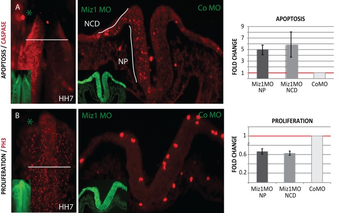
FIGURE 5:
Loss of Miz1 induces apoptosis and defects proliferation in the whole neuroepithelium at HH7. (A) Immunostaining of caspase 3 shows increased apoptosis on the morphant side visible in the whole mount and transverse section of an HH7 embryo. When quantified, Miz1 Mo causes a fivefold increase in apoptosis in the neural plate (NP) and an almost sixfold increase in the neural crest domain (NCD) in the lateral neural folds as compared with the control MO–injected side, which was normalized to 1. (B) Immunostaining of the mitosis marker phospho–histone H3 shows decreased proliferation on the Miz1 MO side. The proliferation in the neural plate, as well as in the neural crest domain, was only 0.6-fold vs. the contralateral side, which was normalized to 1.

