FIGURE 9:
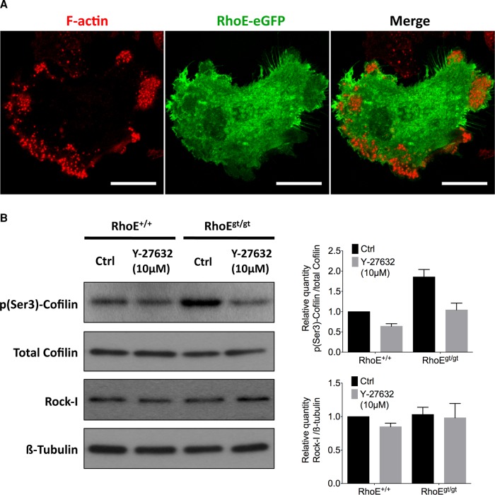
RhoE is cytoplasmic and inhibits Rock-I from phosphorylating cofilin. (A) Micrograph of a day-5 BM-OC expressing RhoE-eGFP (green) 24 h posttransfection and stained for F-actin (red), showing ubiquitous, cytoplasmic localization of RhoE. (B) Immunoblotting of total cofilin, phosphorylated cofilin at Ser-3 (p(Ser-3)-cofilin), Rock-I, and β-tubulin from total cell lysate of day-5 BM-OCs that were serum induced for 2 h with or without 10 μM Y-27632 (a Rock inhibitor) after 2 h of serum starvation. Quantification of protein expression of p(Ser-3)-cofilin normalized to total cofilin reveals 1.86-fold increase in RhoEgt/gt OCs compared with RhoE+/+ OCs without affecting overall Rock-I levels. Diagrams show mean ± SEM from three independent experiments.

