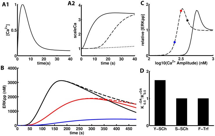
Figure 3. Sensitization of NMDAR Ca2+ triggered ERK activation by dopamine in cultured MSNs.
A1) Unitary ERK activating Ca2+ transient whose amplitude is scaled up by DA-triggered signaling. A2) DA-induced Ca2+-scaling factors through different mechanism: single channel enhancement via tyrosine phosphorylation (ySCh, solid), single channel enhancement via serine/threonine phosphorylation by PKA (sSCh, dashed) and traffic based enhancement by Fyn phosphorylation of NR2B subunits (yTrf, dotted). B) Time course of ERK activation at three different Ca2+ pulse amplitudes in the presence of dopamine. In the dashed curves, the ERK-DUSP negative feedback was turned off. The colors correspond to the dots in panel C. C) The dose-response curve of Ca2+ triggered ERK activation at 8 minutes after dopamine (3 uM) addition (10 minutes in the experimental data) (dashed) is left-shifted relative to the control with no dopamine (solid) for the ySCh mechamism. In both curves ERKpp was normalized relative to the value at 1 uM of Ca2+ amplitude ([ERKpp] = 650 nM). D) Dopamine-triggered sensitization for the three mechanisms expressed as the ratio of the half activating Ca2+ without over with dopamine. Just the ySCh mechanism is sensitizing in these conditions as the other are too slow to boost the early Ca2+ transient.

