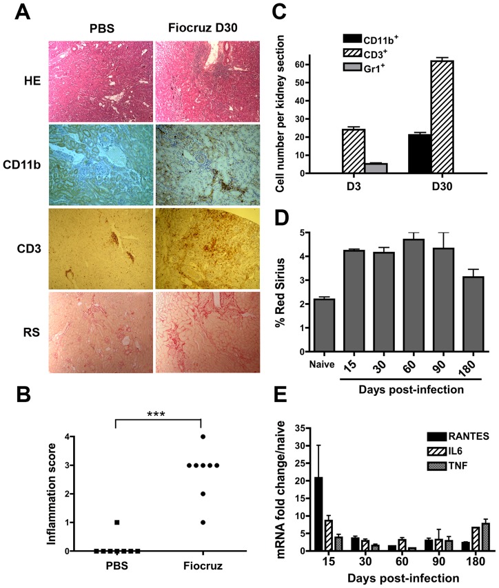
Figure 1. Leptospira infection triggers inflammation and fibrosis in the mouse kidney.
(A) Light microscopy of nodular infiltrates stained with hematoxylin-eosin (HE), infiltrating CD11b+ macrophages and CD3+ T cells and collagen deposition stained with Red Sirius (RS) in kidneys from C57BL/6J mice 30 days (D30) after the inoculation of 2×10⁸ L. interrogans strain Fiocruz. As controls, mice were injected with PBS. Magnification, ×100. (B) Score of kidney inflammation of interstitial nodular infiltrates per surface areas from five different renal tissue sections in control (PBS) and L. interrogans strain Fiocruz infected mice (n = 8 per group). (C) Quantification of the number of CD11b+ macrophages, Gr1+ neutrophils and CD3+ T Lymphocytes per surface area in kidneys from day-3 (D3) and day-30 post-infected mice. (D) Fibrosis quantification by Red Sirius morphometry, expressed as percent of surface area and (E) Inflammation evaluation by mRNA expression of proinflammatory mediators in kidneys of 10 infected mice sacrificed at different time points. Values are means ± SD of counts (C and D) and mRNA quantification (E) from 5 different tissue sections from n = 2 separate mice in each group tested. ***P<0.001.

