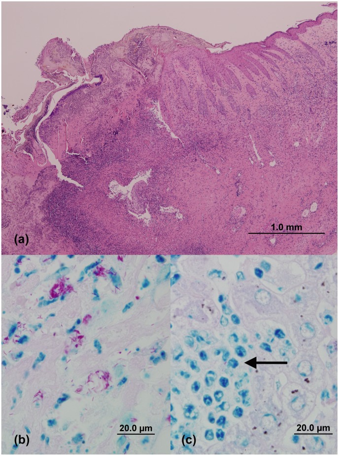
Figure 4. (a): Photomicrograph of a skin lesion obtained from case 16.
The lesion is characterised by proliferative epidermis overlying fibrotic dermal tissue admixed with inflammatory cells, and superficial crust composed of serous exudate and degenerate leukocytes overlying a necrotic base. (H&E stain)) (b): Demonstration of numerous acid-fast bacilli (AFB) in an ulcerated skin lesion of case 16 (modified ZN stain), (c): the liver lesions in this case contained rare AFB, mostly within macrophages (arrow) (modified ZN stain). (Images C. McCowan).

