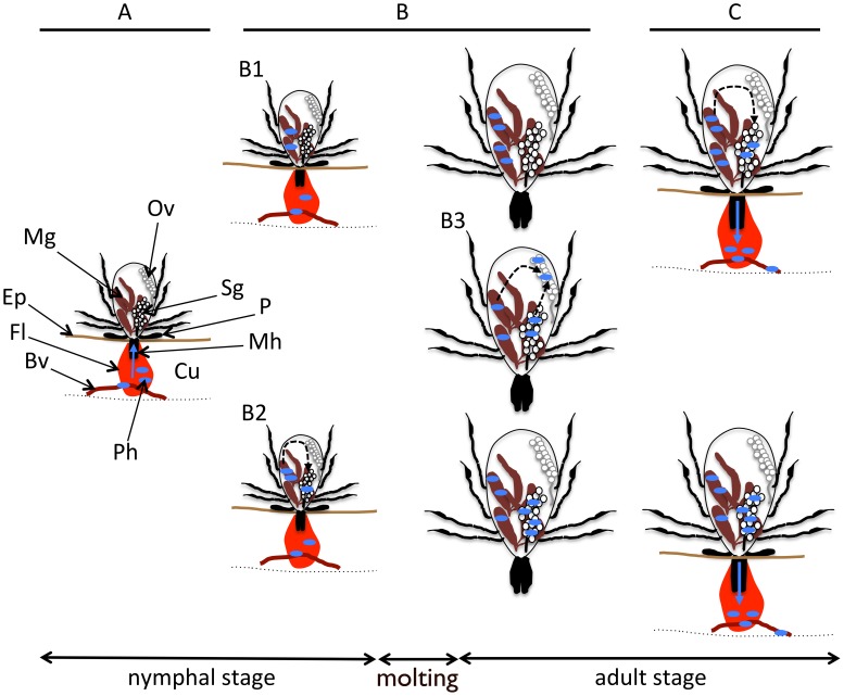
Figure 1. Possible TBP transmission route from an infected host to a new host, via hard ticks.
Note that pathogen multiplication can occur in both the tick midgut or salivary glands, depending on the pathogen. Arrows indicate migrating pathogen pathways. A: Acquisition of TBP by a nymphal stage tick during blood feeding. B: TBP development within the tick; preservation in the tick gut (B1); dissemination into the hemolymph and migration to the salivary glands, which can occur either immediately after acquisition (B2) or after the stimulus of a new blood meal (C); dissemination into the hemolymph and migration to the ovaries (B3), which may or may not occur, and which can lead to transovarial transmission and infection of the succeeding generation. C: TBP transmission from the subsequent adult tick stage to a new vertebrate host during blood feeding; BV: blood vessel; CU: cutis; EP: epidermis; FL: feeding lesion; MG: midgut; MH: mouthparts (chelicera and hypostome); OV: ovaries; P: palp; TBP: tick-borne pathogens; SG: salivary glands. Small blue ovals represent TBP.

