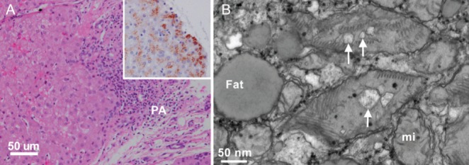
Figure 2).

Light and electron microscopy of liver biopsy. A Periportal area with relatively well-preserved hepatocytes (left) and a portal area (PA) with mild to moderate lymphocytic infiltrate and fibrosis (hematoxylin and eosin stain, bar 50 μm). Insert: granular copper deposits in cytoplasm of hepatocytes (rubeanic acid stain for copper). B High-magnification electron micrograph of hepatocyte cytoplasm containing fat droplets (Fat) and mixture of normal mitochondria (mi) and ‘mega’ mitochondria with dilated mitochondrial cristae (arrows, bar 50 nm)
