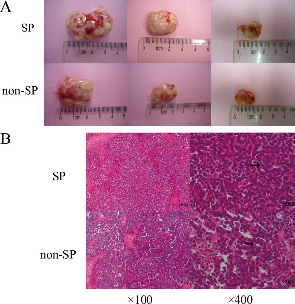
Figure 5.
Tumor formation in NOD/SCID mice and H&E staining result. (A) After inoculated with 1 × 10⁵ (left), 1 × 10⁴ (middle)and 2 × 10³ (right) SP or non-SP cells to NOD/SCID mice, it seemed no statistically significant differences in tumorigenicity, i.e. incidence, latency and growth rate between SP and non-SP cells. (B) Representative H&E stained photomicrographs of SP and non-SP tumors. The tumor resulting from non-SP cell was similar to cervical adenocarcinoma, and it contained duct lumen. As for tumor induced by SP cells, tissue lost their typical characteristics of adenocarcinoma with more obvious cell atypia and fewer duct lumen.

