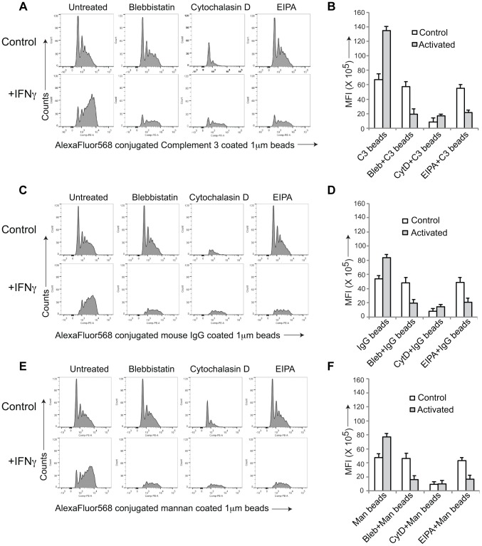
Figure 2. General induction of upon macrophage activation.
AlexaFluor 568 conjugated Complement type 3 coated (A,B), mouse IgG coated (C,D) or mannan-coated 1 µM beads (E,F) were added to resting or activated macrophages in the absence or presence of the indicated inhibitors and analyzed by FACS as described in the legend to Figure S1. Panel B, D and F, depict the mean fluorescence intensity in resting versus activated macrophages obtained for 10,000 cells (data represents average of duplicate samples from three independent experiments).

