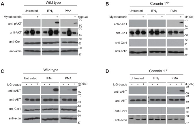
Figure 7. Induction of PI 3 kinase activity in the presence and absence of coronin 1.
A,B. Wild type (A) or coronin 1-deficient (B) macrophages were left untreated or activated with IFN-γ (20 hr) or PMA (4 hrs.) followed by incubation with M. bovis BCG for 0 (−) or 5 (+) min. Cells were lysed and immunoblotted using anti-phospho AKT (Ser473), anti-panAKT, anti-coronin 1 and anti-actin antibodies. The first lanes in each panel represent lysates from cells to which no bacteria were added. C,D. Wild type (C) or coronin 1-deficient (D) macrophages were left untreated or activated with IFN-γ (20 hr) or PMA (4 hrs.) followed by incubation with IgG-coated beads for 0 (−) or 30 (+) min. Cells were lysed and immunoblotted using anti-phosphoAKT (Ser473), anti-panAKT, anti-coronin 1 and anti-actin antibodies. The first lanes in each panel represent lysates from cells to which no beads were added.

