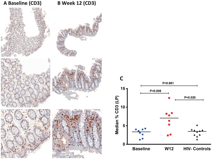
Figure 4. Increase of CD3+ cells in the lamina propria (LP) by immunohistochemistry at week 12 after r-hIL-7 administration.
The percent area of the LP staining for CD3 was evaluated at both baseline, pre-IL-7 (A), and at week 12, post-IL-7, (B) as shown in representative IHC images from one study participant with brown stained cells depicting CD3+ cells at 50× (top panel), 100× (middle panel) and 200× (bottom panel) images. (C) Study participants had a significant increase in CD3+ cells at week 12 compared to baseline (P = 0.008 by paired signed rank test) leading to increased CD3+ surface area compared to surface areas of controls (P = 0.020, Mann-Whitney test). Baseline indicates prior to any drug administration, gut biopsies were performed between 1 and 3 weeks prior to the first (D0) r-hIL-7 injection.

