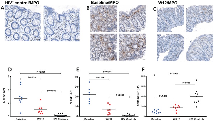
Figure 5. Decreased neutrophil infiltration by myeloperoxidase (MPO) staining, decreased TNF and increased FOXP3 in LP at week 12 after r-hIL-7 administration.
Examples of IHC images of MPO staining in an HIV− control (A), a study participant prior to r-hIL-7 administration (B) and at week 12 after r-hIL-7 (C). Summary results of the percent area of the LP staining for MPO (D) showing a significantly higher number of neutrophils in HIV+ samples than in HIV− controls' (P<0.001) and a decrease at week 12 after r-hIL-7 administration compared to baseline (P = 0.039) that remained different from HIV− controls (P<0.001). The percent area of LP staining for TNF (E) was also higher in HIV+ participants at baseline than among HIV− controls (P<0.001), and decreased significantly after r-hIL-7 (P = 0.016 by Wilcoxon matched pairs rank). In contrast, intracellular FOXP3 staining/mm2 in LP (F) was significantly lower in HIV+ participants than among controls (P<0.001) and increased significantly at week 12 (P = 0.016). Baseline indicates prior to any drug administration, gut biopsies were performed between 1 and 3 weeks prior to the first r-hIL-7 injection.

