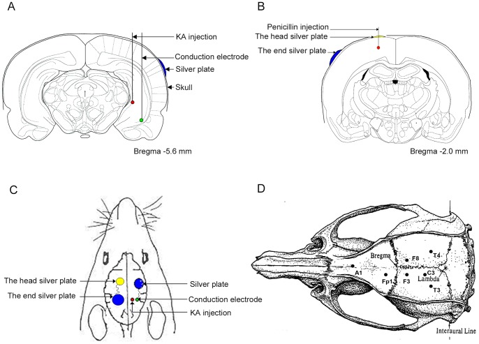
Figure 2. The position of drug injection and electrodes implantation.
(A) and (C): Red, KA injection site, and the tip of the recording electrode for the CA3 region of the right hippocampus; Green, the tip of the conduction electrode; and Blue, the end of the conduction electrode, a silver plate. (B) and (C): Red, penicillin injection site; Yellow, the head end of conduction electrode; and Blue, the tail end of conduction electrode. (D) Diagram of the placement of implanted recording electrodes. The EEG signals were recorded from F4, T4, and T3 electrodes in KA rats, and Fp1, F3 and C3 electrodes in penicillin rats against the reference electrode A1 (adapted from Paxinos & Watson, 1997 [19]).

