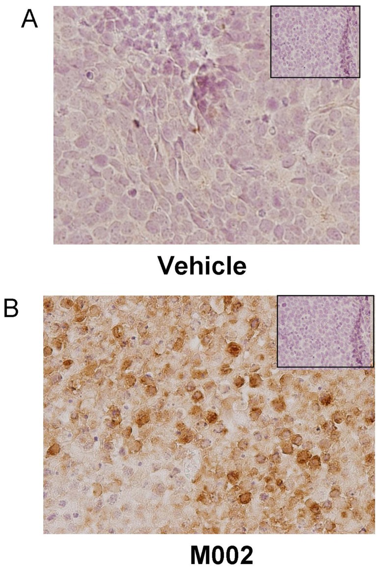
Figure 8. Immunohistochemical staining for HSV in SK-NEP-1 tumor xenografts.
Formalin-fixed, paraffin embedded samples of SK-NEP-1 tumor xenografts (those presented in the data in Figure 5) were stained for HSV using immunohistochemistry, and representative photomicrographs presented. A No HSV was detected by immunohistochemical staining in SK-NEP-1 tumors treated with vehicle alone. Negative controls (rabbit IgG) were included with each run (small box insert). B HSV immunohistochemical staining revealed HSV present in the M002 treated SK-NEP-1 xenografts (brown stain). There was no viral staining seen in the negative controls (rabbit IgG) (small box insert).

