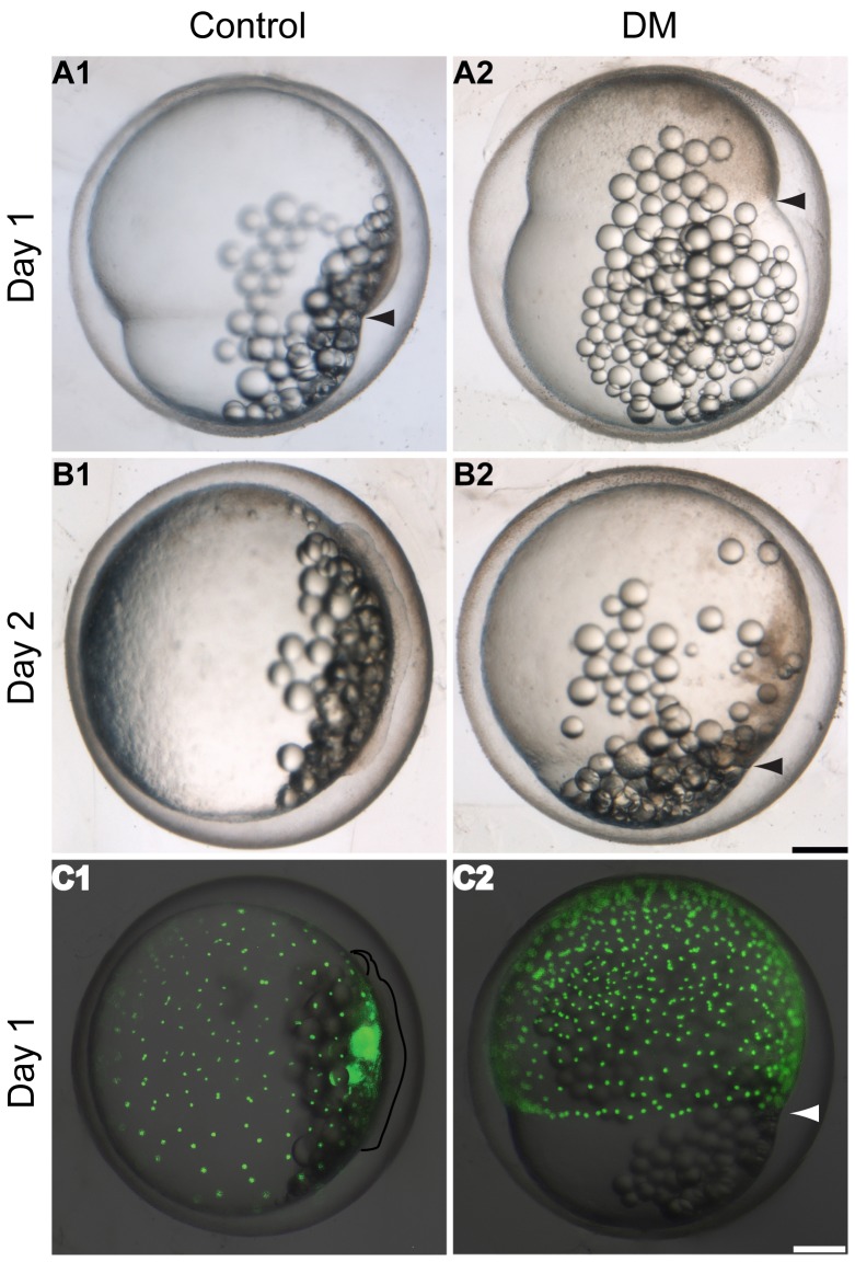
Figure 2. Bmp inhibition delays epiboly progression in K. marmoratus.
Epiboly coverage was recorded at day 1 (A) and day 2 (B) post-fertilization (dpf) in embryos exposed to 100 µM dorsomorphin (DM) at the 32-cell stage. Progression of the yolk syncytial layer (YSL) during gastrulation was assessed via staining of yolk syncytial nuclei (YSN) using Sytox Green. The green fluorescent YSN were observed 1 dpf (C). A1, 2: As control embryos reach c. 70% epiboly (A1 arrowhead, n = 10/10), DM treated embryos are delayed with epiboly covering c. 30% of the yolk (A2 arrowhead, n = 10/10). B1, 2: Controls reach the otic vesicle formation stage (B1, n = 10/10) whilst exposed embryos are lagging behind around 90% epiboly (B2 arrowhead, n = 10/10). C1, 2: Shortly after epiboly closure, control embryos enter the eye formation stage (C1, n = 10/10) (embryo and the eye are outlined) and YSN are spread all over the yolk. On the other hand DM exposed embryos are still mid-epiboly and fluorescent YSN are observed near the blastoderm margin (C2 arrowhead, n = 10/10), demonstrating that YSN are also delayed by inhibition of Bmp signalling. All images are lateral views of the embryos. Scale bars: 250 µm.

