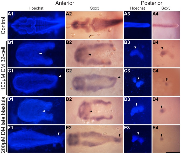
Figure 3. The neural tube is separated in embryos of the splitbody phenotype.
K. marmoratus embryos were exposed to 100 µM dorsomorphin (DM) at the 32-cell stage (B, C), and 200 µM DM at the late blastula stage (D, E) of development. These embryos were then fixed 3 days post-fertilization and used for in situ hybridization using a sox3 probe (stains all neural tissue) (A–E2, A–E4) and Hoechst staining (a blue fluorescent DNA stain) (A–E1, A–E3) in order to examine body contour and split neural tube (A–E1, 2), and the nature of the posterior isolated cell lumps or cell islands (A–E3, 4). A1–4: Control embryo (n = 20/20). B, D: Splitbody individual with an opened end of the body axis and neural tube split (B1, 2 arrowheads n = 19/20, and D1, 2 arrowheads n = 12/20). Splitbody individuals with a closed end, as both strands of the body axis and neural tube join in their most posterior region (C1, 2 arrowheads n = 1/20, and E1, 2 arrowheads n = 8/20). All DM embryos presented here generated cell islands (B-E3 arrowheads) with distinct sox3 positive staining (B–E4 arrowheads). All images are dorsal views of the embryos. Scale bar: 250 µm.

