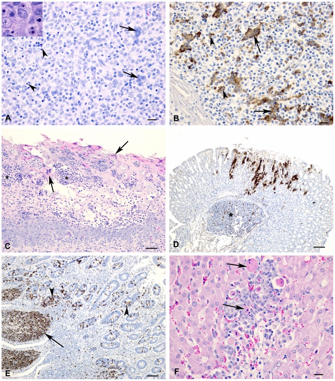
Figure 4. Histology and immunohistochemistry of sheep and goat tissue at varying time points following infection with PPRV (Malig strain).
(A) Section of a lymph node; goat, 6 dpi. Multinucleated syncytial cells (arrows) and degenerating or apoptotic lymphocytes (arrowheads) were observed at 6 and 8 dpi. Inset: Higher magnification showing detail of apoptotic lymphocytes. HE stain, bar = 20 µm (B) Lymph node; goat, 6 dpi. Positive immunostaining using PPRV-specific antibodies in syncytial cells (arrows) and macrophages (arrowheads). Bar = 20 µm. (C) Section of omasum; sheep, 8 dpi. There is necrosis and loss of epithelium with edema, neutrophil infiltration (*) and syncytial cell formation (arrows). HE stain, bar = 50 µm. (D) Abomasum; goat, 8 dpi. Positive immunostaining for PPRV antigen could be detected within the gastric pits and glands as well as in the associated lymphoid tissue (*). Bar = 100 µm. (E) Ileum; sheep, 6 dpi. There is positive immunostaining for PPRV antigen within Peyer’s patches (arrow) as well as crypt epithelial cells (arrowhead). Bar = 50 µm. (F) Liver; sheep, 8 dpi. Focal area of hepatocyte loss with non-suppurative inflammation and degenerating syncytial cells (arrows). HE stain, bar = 50 µm.

