
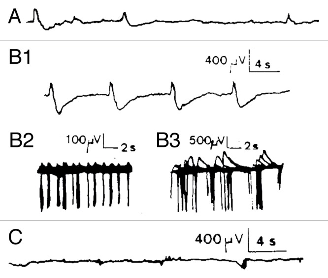
Figure 2. Chemical activation of Kalanchoë Daigremontiana stem tissues. (A) recording of spontaneous spikelets 5 min before FC activation. FC activation responses occurred 7 min after the local FC deposition (3.7 µM, n = 4) between both recording electrodes as trains of −/+ spikes in which the rising phase is 1/3 of the descending one (B1 and B2). This corresponds to classical hyperpolarizations observed in mammalian cells linked to calcium dependent K+ conductance. AP duration ranged from 300–500 msec for the fastest, and up to 2 sec for the slowest, with maximal amplitudes of 3 mV, and separation intervals from 2–16 sec Return to the baseline ranged from 0.25–2.5 sec depending on the duration of the repolarizing phase. The total duration of this burst was 27 min and it was immediately interrupted by application of FCCP 50 µM at the same site (C1, n = 2). However, no restoration was seen, even after 2 h recording, which was not the case for other metabolic inhibitors like 2–4-DNP (100 µM) or low temperature (0°C) that show partial or total recovery, possibly due to the capacity of the tissue to more easily recover from a block of ATP (not shown). Concerning the Material and Methods (electrophysiological set-up, soil culture conditions, temperature and humidity %, stimulating procedure, elimination of interference signals by Faraday cage, correlated methods, use of several kind of electrodes including Ag-AgCl probes, controls made with salt bridges during several hours etc...), please refer to Debono and Bouteau (1992) referenced in 9.