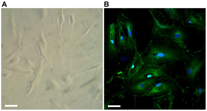
Figure 4. Characterization of ARPC morphology.

(A) Light microscopy image of ARPCs. (B) ARPCs can be differentiated in renal tubular cells, and form junctions as shown by ZO-1 immunostaining (rabbit anti-human ZO-1 polyclonal Ab, green). To-pro-3 counterstains nuclei (blue). Scale bar = 40 µm.
