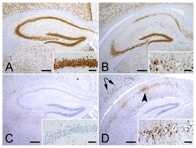
Figure 4. Neuropathology of TMEV infection.
(A) In C57Bl/6 mice NeuN staining demonstrated no degeneration in the hippocampus 12 days post-infection with TMEV. Inset shows a higher magnification of the CA1 region. (B) In Vα14 Tg C57Bl/6 mice NeuN staining revealed neuronal loss in CA1 and CA2 regions of the hippocampus 12 days post-infection with TMEV. (C) On consecutive sections, TMEV protein could not be detected in C57Bl/6 mice, while in the Vα14 Tg C57Bl/6 mice (D) TMEV was readily detectable in the hippocampus (arrowhead) and in the cortex (arrows). Insets show higher magnifications of the CA1 region.

