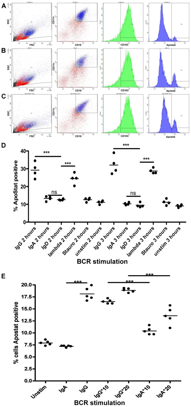
Figure 4. Signalling via BCR using soluble or immobilised anti-sIg antibodies induces apoptosis in IgD−ve mult-HCL.

Tumor cells were stimulated with goat F(ab’)2 anti-Ig antibodies at 20 µg/ml or immobilised mouse anti-Ig using immunobeads at 10 or 20 µg/ml (represented as IgG*, IgA*) for the indicated times at 37°C and early onset apoptosis measured using the Apostat assay. Panels A–C show as an example FC plots from Case 10 (sIgA++, IgG+++, IgM+, lambda+++, with functional BCR isotypes IgG and λ) revealing increased apoptosis (% Apostat positive CD19HICD11cHICD103+ HCs) in response to soluble (Panel B) and immobilised (Panel C) anti-IgG as compared to response to non-functional isotype (anti-sIgA) as control (Panel A). Panel D shows representative data from 1 of 2 replicate experiments using soluble anti-sIg stimuli and increase in apoptosis in response to both functional sIgG and λ mediated signals as compared to a negative control isotype that is not expressed (anti-sIgD). Staurosporin, included as a potential agent for apoptosis, had no measurable apoptotic effect at time of assay. Panel E shows data from 1 of 2 replicate experiments comparing soluble –vs- immobilised anti-sIg stimuli for 3 h (data read-outs shown in Table 2). The data shows that immobilised anti-sIg stimulation also triggers apoptosis (at both 10 and 20 µg/ml) as compared to the immobilised non-functional isotype control (anti-sIgA). Data were analysed in PRISM using Student’s t-test with Welch’s correction. P-values are denoted as: ns>0.05, *0.01–0.05, **0.01–0.001, ***0.001–0.0001, ****<0.0001.
