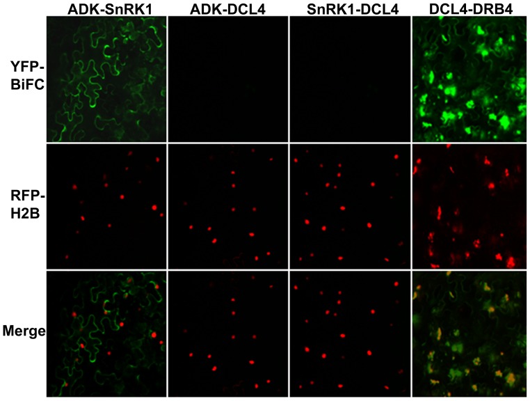
Figure 2. SnRK1 and ADK interact in the cytoplasm.
Constructs expressing full-length SnRK1, ADK, DCL4, or DRB4 proteins fused to the N- or C-terminal portion of YFP were delivered by agroinfiltration to N. benthamiana leaves. Images were captured at 40x magnification 48 h post-infiltration using a confocal microscope. Representative images of epidermal cells, which have a characteristic irregular shape and a large vacuole that restricts the cytoplasm to the cell periphery, are shown. Histone H2B fused to RFP (RFP-H2B) is a marker for the nucleus.

