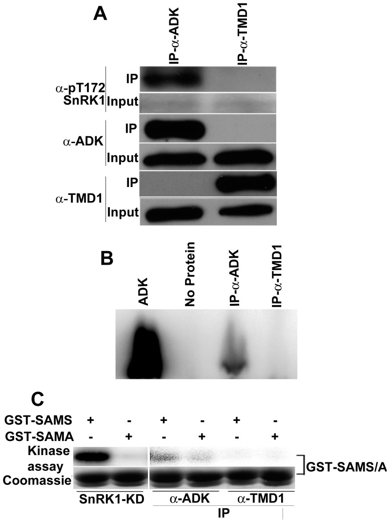
Figure 3. SnRK1 co-immunoprecipitates with ADK.
(A) Immunoprecipitation (IP) and immunodetection were performed with ADK antibody (α-ADK) and TMD1 antibody (α-TMD1). Activated SnRK1 was detected using an antibody raised against phosphorylated AMPK (α-pT172). (B) Immunoprecipitates obtained with α-ADK or α-TMD1 were incubated with 1 µM adenosine and γ32P-ATP. Controls contained purified ADK or no added protein. Reaction mixtures were resolved by thin layer chromatography and labeled AMP was detected using a phosphor-imager. (C) Immunoprecipitates obtained with α-ADK or α-TMD1 were incubated with 3 µg of GST-SAMS or GST-SAMA and γ32P-ATP. Assays with purified SnRK1-KD were included as controls. Samples were fractionated on SDS-PAGE gels and exposed to a phosphor-imager for 2 h (SnRK1-KD controls) or 2 days (immunoprecipitate samples). GST-SAMS and GST-SAMA were monitored by Coomassie stain.

