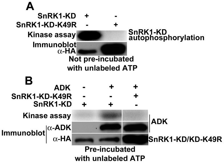
Figure 5. ADK is phosphorylated by SnRK1 in vitro.
Kinase assays were conducted using γ32P-ATP with SnRK1-KD or SnRK1-KD-K49R either alone or with ADK. With the exception of autophosphorylation assays, SnRK1-KD and SnRK1-KD-K49R were pre-incubated with unlabeled ATP to obscure autophosphorylation. Samples were fractioned by SDS-PAGE and signals detected using a phosphor-imager. Immunoblots were probed with anti-ADK to detect ADK, or with anti-HA to detect SnRK1-KD or SnRK1-KD-K49R. (A) Kinase assays with SnRK1-KD (10 ng) and SnRK1-KD-K49R (30 ng) were performed without pre-incubation with unlabeled ATP. (B) ADK protein (3 µg) was incubated with SnRK1-KD (10 ng) or SnRK1-KD-K49R (30 ng). Note that SnRK1-KD autophosphorylation (in the lane lacking ADK) was nearly undetectable due to pre-incubation of SnRK1 with unlabeled ATP. The same pre-incubated SnRK1-KD and SnRK1-KD-K49R preparations were used to perform the GST-SAMS phosphorylation experiment shown in Figure 1C. Activity data are presented in Table S3.

